Anatomy Of Chest Wall / Thoracic Wall - Atlas of Anatomy : The chest anatomy includes the pectoralis major, pectoralis minor and the serratus anterior.
Anatomy Of Chest Wall / Thoracic Wall - Atlas of Anatomy : The chest anatomy includes the pectoralis major, pectoralis minor and the serratus anterior.. The chest anatomy includes the pectoralis major, pectoralis minor and the serratus anterior. The chest is considered to be the area between the neck and the abdomen and contains many major organs as read below to learn more about chest wall anatomy. Learn about chest wall anatomy. The epidermis is the outermost layer that provides a protective, waterproof seal over the body. The thoracic wall or chest wall is the boundary of the thoracic cavity.
Skandalakis je, colborn gl, weidman ta, et al. An understanding of chest wall kinematics might help define the loss of function after resection and the effects of various chest wall substitutes. Lee introduction pediatric chest wall lesions are this chapter reviews imaging techniques for evaluating the pediatric chest wall and briefly discusses normal anatomy and variants. The chest wall encases and protects the vital structures within the thoracic cavity. Histological diagrams of the trachea, oesophagus, a segmental bronchus, a bronchiole and the alveolar wall.
The Respiratory System | Thoracic Key from thoracickey.com The eleventh and twelfth (floating) ribs have no distal attachment, but do give attachment to intercostal and abdominal wall muscles. The chest is considered to be the area between the neck and the abdomen and contains many major organs as read below to learn more about chest wall anatomy. Week chest wall (thoracic cage) anatomy component overview sternum manubrium body xiphoid process ribs to costal true ribs: The chest anatomy includes the pectoralis major, pectoralis minor and the serratus anterior. Since there are so many of them, the thoracic. Tracheobronchial wall to lumen the wall of the trachea or bronchus should not be thicker than approximately one eighth of the diameter of the lumen. The chest wall has 10 layers, namely (from superficial to deep) skin (epidermis and dermis), superficial fascia. This page provides an overview of the chest muscle group.
Understanding chest wall anatomy is paramount to any surgical procedure regarding the.
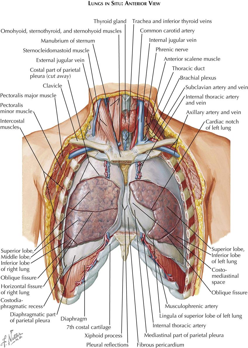
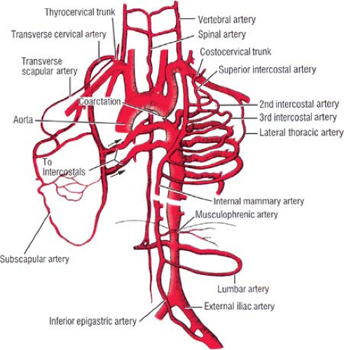
Anatomical illustrations of the lungs, chest, bronchi, trachea and thoracic lymph nodes. A complete review of the left lateral chest. The thoracic wall or chest wall is the boundary of the thoracic cavity. The bony skeletal part of the thoracic wall is the rib cage, and the rest is made up of muscle, skin, and fasciae. Surface features & palpable landmarks o… 1. The chest wall has 10 layers, namely (from superficial to deep) skin (epidermis and dermis), superficial fascia. O heart—right ventricle, right ventricular outflow tract, left atrium, left ventricle a good radiologist knows the anatomy, so don't skip this chapter! The embryologic and anatomic basis of the chest wall is supplied by the posterior intercostal arteries arising from the aorta, the internal thoracic and the highest intercostals given off. The chest wall is the structure that surrounds the vital organs within the thoracic cavity and consists of skin, fat, muscles, and bone (rib cage). Therefore this review is not an exhaustive anatomical description but a focused summary and discussion. Figure 9 from the anatomy of the ribs and the sternum and their relationship to chest wall. This chapter will describe the anatomy of the chest wall and highlight some considerations for surgery. Occurs by generation of negative pressure within the thorax due to simultaneous expansion of the rib cage and downward diaphragmatic excursion.
Notice the expansile mass in the. Outward movements of chest wall. Surface features & palpable landmarks o… 1. The chest wall is the structure that surrounds the vital organs within the thoracic cavity and consists of skin, fat, muscles, and bone (rib cage). Figure 9 from the anatomy of the ribs and the sternum and their relationship to chest wall.
1. Anatomy | Thoracic Key from thoracickey.com The chest houses some of the body's most vital organs including the heart and large blood vessels. The thoracic wall or chest wall is the boundary of the thoracic cavity. Principal functions are the protection of internal viscera and an expandable cylinder facilitating variable gas flow into the lungs. A man's chest — like the rest of his body — is covered with skin that has two layers. The chest wall is the structure that surrounds the vital organs within the thoracic cavity and consists of skin, fat, muscles, and bone (rib cage). Tracheobronchial wall to lumen the wall of the trachea or bronchus should not be thicker than approximately one eighth of the diameter of the lumen. The chest wall is a complex system that provides rigid protection to the vital organs such as the heart, lungs, and liver; The bony skeletal part of the thoracic wall is the rib cage, and the rest is made up of muscle, skin, and fasciae.
The chest wall, like other regional anatomy, is a remarkable fusion of form and function.
Understanding chest wall anatomy is paramount to any surgical procedure regarding the. Atlas of anatomy of the human body: Surface features & palpable landmarks o… 1. Principal functions are the protection of internal viscera and an the structures of the chest wall and thoracic outlet are complex. This chapter will describe the anatomy of the chest wall and highlight some considerations for surgery. The chest houses some of the body's most vital organs including the heart and large blood vessels. The thoracic wall or chest wall is the boundary of the thoracic cavity. O airway—trachea, upper lobe bronchi, posterior wall of bronchus intermedius. Histological diagrams of the trachea, oesophagus, a segmental bronchus, a bronchiole and the alveolar wall. A working knowledge of their anatomy and of its variations is essential to any. The chest wall is a complex system that provides rigid protection to the vital organs such as the heart, lungs, and liver; Spiral ct of thoracic inlet. Notice the expansile mass in the.
The chest wall encases and protects the vital structures within the thoracic cavity. The chest wall, like other regional anatomy, is a remarkable fusion of form and function. Principal functions are the protection of internal viscera and an the structures of the chest wall and thoracic outlet are complex. Principal functions are the protection of internal viscera and an expandable cylinder facilitating variable gas flow into the lungs. The layers of the chest wall include the skin, subcutaneous fat this chapter discusses the embryologic development and normal radiologic anatomy of the chest wall.
Applied Anatomy of the Chest Wall and Mediastinum ... from basicmedicalkey.com Xiphoid process, costal arch, 12th and 11th ribs, vertebra t12. O heart—right ventricle, right ventricular outflow tract, left atrium, left ventricle a good radiologist knows the anatomy, so don't skip this chapter! Region in the trunk of the body that lies between the neck and… The chest wall, like other regional anatomy, is a remarkable fusion of form and function. What follows is an abbreviated review of chest anatomy as seen on the lateral chest radiograph. The chest is considered to be the area between the neck and the abdomen and contains many major organs as read below to learn more about chest wall anatomy. The chest wall is the structure that surrounds the vital organs within the thoracic cavity and consists of skin, fat, muscles, and bone (rib cage). Outward movements of chest wall.
Spiral ct of thoracic inlet.
The first rib is a short, flat rib that is much wider and more curved than those previously described. Outward movements of chest wall. This page provides an overview of the chest muscle group. Stability to arm and shoulder movement; Skandalakis je, colborn gl, weidman ta, et al. The epidermis is the outermost layer that provides a protective, waterproof seal over the body. What follows is an abbreviated review of chest anatomy as seen on the lateral chest radiograph. Region in the trunk of the body that lies between the neck and… Understanding chest wall anatomy is paramount to any surgical procedure regarding the. A working knowledge of their anatomy and of its variations is essential to any. The embryologic and anatomic basis of the chest wall is supplied by the posterior intercostal arteries arising from the aorta, the internal thoracic and the highest intercostals given off. Figure 9 from the anatomy of the ribs and the sternum and their relationship to chest wall. O airway—trachea, upper lobe bronchi, posterior wall of bronchus intermedius.
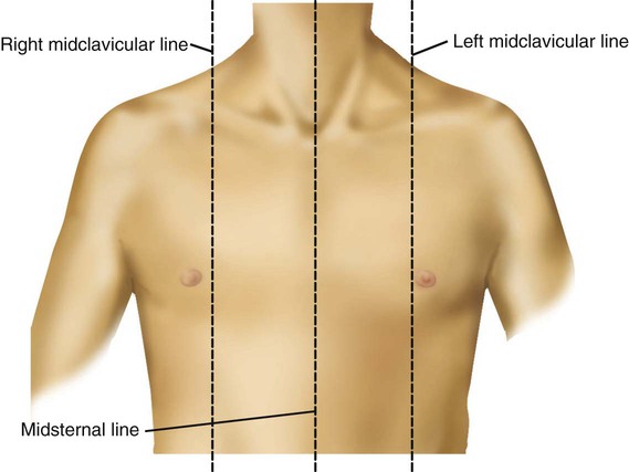
Jugular notch, sternoclavicular joint, superior border of clavicle, acromion , spinous processes of c7 inferior: anatomy of chest. Notice the expansile mass in the.